Protein Electrophoresis For M Band
The serum protein electrophoresis SPEP test measures specific proteins in the blood to help identify some diseases. In other cases patients may have non-specific symptoms meaning the discovery is more unexpected.
Serum Protein Electrophoresis Gel Proteinwalls
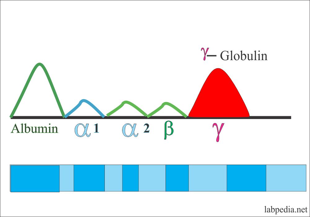
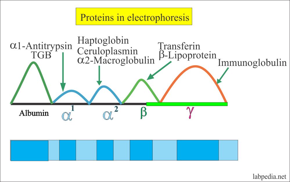
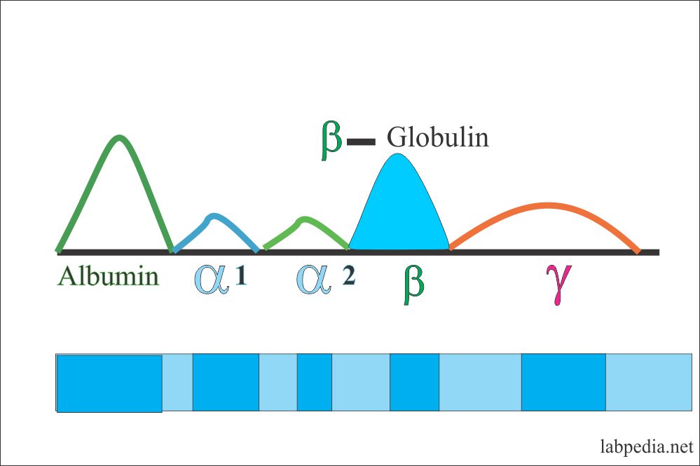
Test results for each protein group are given as a percentage of the total amount of serum protein.

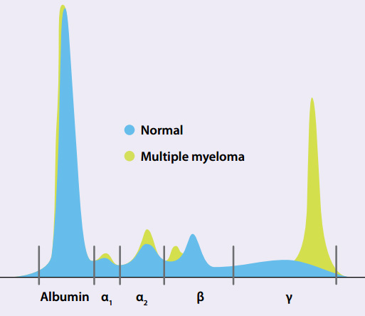
Protein electrophoresis for m band. 15052020 If the serum M-protein spike is 15 to 25 g per dL it is important to perform nephelometry to quantify the immunoglobulins present and to obtain a 24-hour urine collection for electrophoresis. 08042021 To evaluate M-protein serum protein electrophoresis SPEP is used where a single band known as M-band is seen. A characteristic monoclonal band M-spike is often found on serum protein electrophoresis SPE in the gamma-globulin region and more rarely in the beta or alpha-2 regions.
However in rare entities like biclonal gammopathy two M-bands appear simultaneously at different positions on SPEP which may be attributed to the clonal expansion of two different neoplastic cell lines. If the M band or paraprotein is observed in serum protein electrophoresis the following steps are performed. The finding of an M-spike restricted migration or hypogammaglobulinemic SPE pattern is suggestive of a possible monoclonal protein.
The M-protein can be evaluated by serum protein electrophoresis SPEP which yields a single discrete band M-band usually in the γ-globulin region. To obtain the actual amount of each fraction a test that measures the total serum protein must also be done. This band is usually seen in the gamma globulin region.
21022019 To find these identical M proteins your doctor might run a blood test called serum protein electrophoresis SPEP. Abnormal Protein Band 1 Optimal Result. That technique also has enhanced our ability to detect genetic variants such as α 1-antitrypsin inhibitor deficiencies PiZZ and PiSZ as well as benign variants.
Serum protein bands monoclonal gammopathy will sometimes be found following serum protein electrophoresis in patients presenting with classic signs or symptoms of multiple myeloma eg. 01082019 Serum and Urine Protein Electrophoresis SPEP and UPEP Serum protein electrophoresis SPEP is a test that measures the amount of heavy chain monoclonal protein made by myeloma cells. Protein electrophoresis is a test that measures specific proteins in the blood.
Serum protein electrophoresis is most commonly ordered when multiple myeloma is suspected and observation of a monoclonal band M band paraprotein indicates that monoclonal gammopathy may be present in the patient. 0 - 0 gdL. It involves placing a sample of.
Protein electrophoresis m band protein electrophoresis method protein electrophoresis methods and protocols protein electrophoresis m spike 1 protein electrophoresis machine protein electrophoresis meaning protein electrophoresis mayo clinic protein electrophoresis methods and protocols pdf protein electrophoresis medscape protein electrophoresis. The test separates proteins in the blood based on their electrical charge. When an abnormal protein band or peak is detected additional tests are done to identify the type of protein immunotyping.
During discontinuous polyacrylamide gel electrophoresis the isolated protein separates into three bands which can be identified as two separate components A B and a complex of the two. 01121972 M band protein can be specifically extracted from fresh chicken breast muscle myofibrils suspended in 5 mM Tris-HCl pH 80. SPEP separates all the proteins in the blood according to their electrical charge.
Electrophoresis also permits quantitation of M proteins. These all separate the proteins into distinct bands or fractions. 10032015 Serum protein electrophoresis SPE is commonly used as a diagnostic tool to screen for plasma cell dyscrasias including multiple myeloma macroglobulinemia and amyloidosis 1.
The presence of M proteins can be a sign of a type of cancer called myeloma or multiple myeloma. Improved resolution especially of protein bands has resulted in better detection of bisalbuminemia by laboratories using capillary zone electrophoresis. The protein electrophoresis test is often used to find abnormal substances called M proteins.
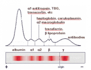
Serum protein electrophoresis SPE separates proteins into multiple bands using an electrical field and can be done in various media including cellulose acetate largely replaced agarose gel or liquid within a capillary tube capillary zone. Screening by SPE is then commonly followed by immunofixation electrophoresis IFE to confirm and classify the specific paraprotein present IgG IgA IgM IgD or IgE. -A characteristic monoclonal band M-spike is often found on serum protein electrophoresis SPE in the gamma globulin region and more rarely in the beta or alpha-2 regions.
Protein electrophoresis of the serum and urine detects M protein as a narrow peak like a church spire on the densitometer tracing or as a dense discrete band on agarose gel Fig. Urine protein electrophoresis or UPEP does the same thing for proteins. Possible monoclonal protein M-protein present.
Learn more at Types of Myeloma.

Serum Protein Electrophoresis A Gel Picture Showing Two Distinct Download Scientific Diagram
Protein Serum Electrophoresis Total Protein Albumin And Globulin Labpedia Net
Protein Serum Electrophoresis Total Protein Albumin And Globulin Labpedia Net
Interpreting Serum Protein Electrophoresis Spe Patterns Bcit News

Immunoglobulin A Gammopathy On Serum Electrophoresis A Diagnostic Conundrum Bansal F Bhagat P Srinivasan Vk Chhabra S Gupta P Indian J Pathol Microbiol
Making Sense Of Serum Protein Bands Best Tests July 2011

Understanding And Interpreting The Serum Protein Electrophoresis American Family Physician
Protein Serum Electrophoresis Total Protein Albumin And Globulin Labpedia Net

Multiple Myeloma Recognition And Management American Family Physician

Post a Comment for "Protein Electrophoresis For M Band"