Formation of a protein corona was characterized by means of dynamic light scattering z potential and liquid chromatographymass spectrometry. 13 nm to the nanoparticle diameter.
Overview Of Key Principles Of Dynamic Light Scattering
λ 20 15 nm A 2 second virial coefficient Parameters derived.

Protein corona dynamic light scattering. Hydrodynamic diameter HD with dynamic light scattering DLS Figure 1C. The zeta potentials of CeO. Methods and techniques for students.
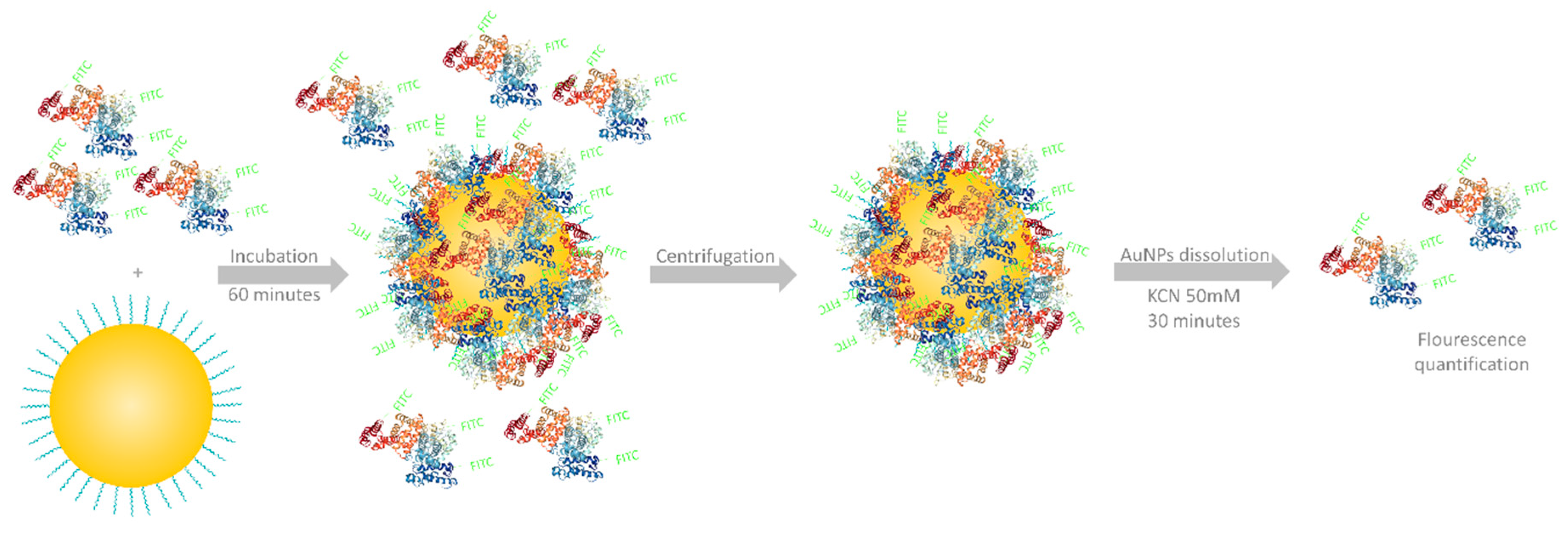
A total of 288 different human serum proteins were identified as shown in the heat map Figure S2. Molar Mass weight-average accuracy 5 rg212 root mean square radii for rg212. 21072013 The protein corona transition from its early dynamic state to the later more stable corona was evaluated using mass spectrometry.
We found that the serum protein corona added 30. The NP diameter was 224. Although dynamic scattering is in principle capable of distinguishing whether a protein is a monomer or dimer it is much less accurate for distinguishing small oligomers than is classical light scattering or sedimentation velocity.
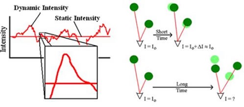
22 nm measured by scanning transmission electron microscopy STEM. Dynamic light scattering DLS analyses are routinely used in biology laboratories to detect aggregates in macromolecular solutions to determine the size of proteins nucleic acids and complexes or to monitor the binding of ligands. The angular dependency of the inverse.
Combination of dynamic light scattering and isothermal titration calorimetry. Of soft and hard corona proteins The particle size distribution of the nanoplastics was first eval-uated with dynamic light scattering DLS for each nanoplastic. During the last two decades the panel of methods used to study the structure and function of proteins and nucleic acids has grown continuously.
07092015 The structure and composition of the liposome-protein corona as determined by dynamic light scattering electrophoretic light scattering and liquid chromatography tandem mass spectrometry were found to be dependent on the incubation protocol. Towards a unified biophysical characterization platform. 15082019 The liposomeprotein corona is a dynamic interface that regulates the interaction of liposomes with the physiological environment.
Table I presents the summary of hydrodynamic diameter and poly-dispersity index PDI all of which suggest that the nanoplastics are moderately monodisperse PDI01 or lower. The advantage of using dynamic scattering is the possibility to analyze samples. Time-averaged intensity of scattered light Dynamic quasielastic fluctuation of intensity of scattered light with time Light Scattering Experiments Parameters derived.
To evaluate the effect of a protein corona on stem cell labeling human. The NPs were incubated in BALf to examine the formation of the protein corona and its influence on NP agglomerate size and zeta potential. 2 nm monitored by multi angle dynamic light scattering attributed to the hard protein corona around nanoparticles.
Combining dynamic light scattering and Raman spectroscopy. Light scattering protein temperature viscosity. The protein composition of this layer was profiled using label-free LC-MS MS.
There is a size increase of 19. The cyclic PEOXA brushes generate NP shells that are denser and more compact than their linear counterparts entirely preventing the formation of a protein corona as well as aggregation even when the lower critical solution temperature of PEOXA in a. Use of DLS combined with Raman spectroscopy to aid in the understanding of the colloidal stability of biotherapeutic formulations with particular regard to the onset of protein aggregation events.
Changes in hydrodynamic diameter and agglomeration kinetics were studied using dynamic light scattering DLS. Iron uptake was evaluated with 339-diaminobenzidine Prussian blue staining lysosomal staining and inductively coupled plasma spectrometry. As four soluble proteins and a ribonucleoprotein assembly purified and characterized by students in the frame of their master degree.
The multiple washing steps removed unbound and loosely bound proteins from the NPs. Protein analysis by dynamic light scattering. In the scope of DLS temporal fluctuations are usually analyzed by means of the intensity or photon auto-correlation function also known as photon correlation spectroscopy or quasi-elastic light scattering.
At 37 C by dynamic light scattering DLS using a Malvern ZetaSizer Nano Westborough MA. 13012020 dynamic light scattering DLS tracked the increase in protein- PNP hydrodynamic radius solution absorbance confirmed proteinPNP association - followed byremoval of unbound protein and quantifying eluted protein verified full desorption of the PNP protein corona. Dynamic light scattering DLS is a technique in physics that can be used to determine the size distribution profile of small particles in suspension or polymers in solution.
19122016 Indeed particle tracking analysis and dynamic light scattering did not show any increase but instead showed a small decrease in the size of particles after binding of serum proteins.
Protein Corona Evaluation A Acrylamide Sds Page Electrophoresis Gel Download Scientific Diagram
![]()
A Dynamic Light Scattering And B Nanoparticle Tracking Analysis Nta Download Scientific Diagram
1
Applied Sciences Free Full Text Quantification Of The Pegylated Gold Nanoparticles Protein Corona Influence On Nanoparticle Size And Surface Chemistry Html

Analysis Of Temporally Evolved Nanoparticle Protein Corona Highlighted The Potential Ability Of Gold Nanoparticles To Stably Interact With Proteins And Influence The Major Biochemical Pathways In Brassica Juncea Sciencedirect

A Representative Dynamic Light Scattering Dls Intensity Download Scientific Diagram

The Main Techniques Used To Study Protein Corona Features Download Scientific Diagram

Dynamic Light Scattering Dls Results Showing The Increase In The Size Download Scientific Diagram

Binding Of Fetal Bovine Serum Fbs Proteins To Nanoparticle Surfaces Download Scientific Diagram


0 comments:
Post a Comment